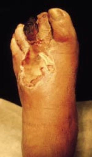
Peripheral artery disease (PAD)
Last edited on : 22/09/2024
Peripheral Artery Disease (PAD) is defined as the presence of progressive stenoses (narrowing) of the peripheral arteries in the lower limbs leading to a reduction in peripheral blood flow. Symptoms appear when collateral circulation becomes insufficient to meet cellular oxygen demands (exercise or rest ischemia, trophic disorders, risk of acute ischemia).
Its prevalence is 1% before age 50 and 7% after age 60. The sex ratio is 3 men to 1 woman before age 65 and equal thereafter.
Potential causes are numerous but are generally those of atherosclerosis.
PAD is usually benign in itself (critical ischemia affecting only 1% of those over 55), but is associated with very poor prognosis conditions: coronary artery disease (50 to 75%), carotid and/or vertebral stenosis (10%), abdominal aortic aneurysm (10%), strokes, etc.
Etiologies and Clinical Forms
- Atherosclerosis: ~70% of cases
- Elderly patients, slow progression
- Localized occlusions, unilateral or bilateral involvement
- Extensive gangrene
- Most affected vessels: aorta, iliac, femoral arteries
- Diabetic Artery Disease: ~20% of cases, generally more severe and more distal
- Younger patients, rapid progression
- Multisegmental occlusions, bilateral involvement
- Frequent distal trophic lesions (++ toes)
- Most affected vessels: anterior and posterior tibial arteries, peroneal, pedal, arterioles
- Arteriosclerosis (senile arteritis = physiological aging): ~5%
- Embolic Artery Disease
- Look for small purpuric spots on the feet, a blue toe, livedo. Such manifestations associated with pulses still present at the ankles should prompt angiography of the entire arterial tree and an assessment for purpura (prioritize ruling out infectious endocarditis).
- Vasculitis
- To be considered in young individuals or in cases with systemic signs or unexplained inflammatory syndrome
- Takayasu's disease, Buerger's disease, polyarteritis nodosa, systemic lupus erythematosus, Horton’s disease, etc.
- Miscellaneous
- To be considered in young individuals or if other suggestive signs are present
- Hemostatic disorders, homocystinuria, etc.
The risk factors are primarily those of atherosclerosis: smoking, diabetes, men > 55 years or women > 65 years, dyslipidemia, hypertension, physical inactivity, obesity, other cardiovascular history.
Clinical and Evolution

Diagnosis is primarily based on anamnesis, with a lesser degree on clinical examination. Complementary exams are mainly used to grade the extent of the disease.
Anamnesis
- Intermittent claudication = the cardinal symptom = pain (or weakness or "cramps") in a limb (intensity and location variable, ++ in the calf) occurring with exertion. Often occurs after the same walking distance or type of effort and subsides within minutes upon stopping.
- Rest pain, relieved by dangling the legs
- Any trophic changes?
- History of diabetes? Cardiovascular diseases? Risk factors (high blood pressure, smoking, etc.)?
Clinical Examination
- Inspection: signs of arterial insufficiency (muscle atrophy, sparse hair, dry and brittle nails, dry and cracked skin, pale and cold feet, cyanosis, painful ulcers, etc.), signs of diabetic foot, etc.
- Comparative palpation of femoral, popliteal, pedial, and posterior tibial pulses. Absent or diminished? Induration or aneurysmal dilation?
- Arterial auscultation of the aorta up to the knees: bruit (stenosis)?
Evolution
- Among those > 55 years:
- 10 to 20% have asymptomatic PAD (incidental discovery, prognostic significance debated)
- Among symptomatic patients:
- 1% have critical ischemia at diagnosis
- 30% mortality at 5 years (with 75% being cardiovascular in origin)
- 70 to 80% will remain stable or improve
- 20 to 30% will progress and require endovascular or surgical revascularization
- 10% will progress to critical ischemia requiring amputation
Complementary Examinations
Biology
- Check for risk factors: glycated hemoglobin, lipids, uric acid, blood count, fibrinogen, homocysteine, CRP-ESR.
Measurement of Perfusion Pressures and Ankle-Brachial Index (ABI)
ABI (measured with Doppler) = Ankle systolic pressure / Arm systolic pressure (bilateral measurements, use the highest value) :
- > 1.3: Mediacalcinosis
- 0.9-1.3: Normal
- 0.41-0.9: Mild to moderate PAD
- < 0.4: Severe PAD
Note that mediacalcinosis is the main differential diagnosis for PAD with reduced or absent peripheral pulses. It can, however, coexist with true PAD and make ABI interpretation difficult.
Hyperemia Test
- In case of diagnostic doubt, the patient can be asked to perform muscle exercise (treadmill/stair stepper) → normally, the tibial pressure increases before returning to normal. In PAD, it decreases.
Transcutaneous Oxygen Pressure
- Alternative if the measured perfusion pressures by Doppler are unreliable (medial calcinosis at ABI but high clinical suspicion of PAD). Normal values on the dorsal side of the foot are 40 to 80 mmHg.
Doppler Ultrasound of Lower Limbs, Carotids, and Abdominal Aorta
Arteriographic Exams
An arteriographic exam is essential if an intervention is considered. It provides the best anatomical information but does not replace the previously mentioned non-invasive exams (no functional assessment).
- Angio-MRI
- Angio-CT Scan
- Conventional Aortography : remains the GOLD standard, although angio-MRI/CT scans tend to replace it, and allows for possible simultaneous treatment. Essential in case of suspected embolic origin.
Fontaine Classification
| Grade | Criteria | |
| I | Asymptomatic lesions | |
| II | Intermittent claudication, stress ischemia | |
| IIa | Walking perimeter > 200 m | |
| IIb | Walking perimeter < 200 m | |
| III | Resting ischemia, decubitus pain | |
| IIIa | Psyst ankle > 50 mmHg | |
| IIIb | Psyst ankle < 50 mmHg (critical ischemia level) | |
| IV | Trophic disorders: ulcerations, gangrene,...
(! "false grade IV": plantar perforation, venous stasis,...!) | |
Differential Diagnoses
- Variable depending on clinical presentation:
- Intermittent claudication → neurogenic claudication due to lumbar stenosis (pain often less intense, accompanied by paresthesias, relieved by flexion of the spine and aggravated by its extension, no trophic disorders), venous claudication (occurs in the context of major phlebitis sequelae, tends to improve with continued walking)?
- Trophic disorders → angioneurotic dermatitis (extensive skin infarcts, ++ elderly hypertensive and/or diabetic patients, very good healing after necrosis excision without revascularization)
- Young patients → Buerger's disease (significant smoking, Raynaud's phenomenon, Allen's test +, etc.), trapped popliteal artery syndrome (extrinsic compression → stenosis, post-stenotic aneurysm, and embolic migrations mimicking leg artery disease), aortic coarctation, fibromuscular dysplasia, endofibrosis, adventitial cysts, etc.?
- Inflammatory context: Horton’s disease? Takayasu’s arteritis? Polyarteritis nodosa (PAN)?
Therapeutic Management - Treatments
Except for rare etiologies (vasculitis, embolic diseases, etc.) requiring specific management, treatment generally involves managing risk factors, anti-aggregation therapy, and possible revascularization.
Managing Risk Factors and Medical Care
- Smoking cessation, correcting metabolic disorders, physical reconditioning (> 3 x 30 minutes/week, ++ walking until the onset of sub-maximal pain, for > 6 months), etc.
- Anti-aggregation: acetylsalicylic acid 80 to 160 mg/day. Does not improve PAD much but prevents associated cardiovascular manifestations.
- The use of ticlopidine, dipyridamole, or clopidogrel is debated.
- Statins (secondary prevention + stabilization of atherosclerotic plaques).
- Anti-hypertensive treatment if needed.
- Pain management.
- The use of vasodilators is debated. They are mainly used in critical ischemia.
- Discuss the use of prostaglandins in severe stages where intervention is not possible.
- Local care of trophic disorders, hypercaloric diet, antibiotics if secondary infections occur.
- β-blockers are traditionally contraindicated, but no evidence-based medicine (EBM) shows a deleterious role; use if necessary (coronary artery disease, heart failure, etc.).
- In diabetes, aim for HbA1c < 6.5%.
Revascularization Techniques
Certain indication in cases of critical ischemia (from stage IIIb). To be discussed for stages IIb and IIIa. The choice between surgery and endovascular treatment depends on lesion anatomy and the expertise of the reference center.
- Endoluminal angioplasty with balloon ± Dacron stent grafts. Higher risk of restenosis with distal intervention. Most commonly used treatment for short lesions.
- Surgery:
- Endarterectomy
- Vein or Dacron bypasses. Most commonly used technique for diffuse lesions.
- Lumbar sympathectomy: may have an effect on pain, rarely indicated.
- Management of other vascular lesions identified during evaluation if necessary (consider endarterectomy for carotid stenoses > 60% if the patient’s life expectancy is > 5 years, consider stenting of vertebral stenoses > 50% in case of recurrent strokes despite optimal medical treatment, management of possible aortic aneurysms, etc.).
Amputations
To be considered in cases of distal gangrene.
Bibliography
Berger JS et al., Overview of lower extremity peripheral artery disease, UpToDate, 2022
Davis MG et al., Approach to revascularization for claudication due to peripheral artery disease, UpToDate, 2022
Davis MG et al., Management of claudication due to peripheral artery disease, UpToDate, 2022
EMC, Traité d'Angéiologie, 2018
Harris L et al., Peripheral artery disease: Prevalence and risk factors, UpToDate, 2022
Neschis DG et al., Lower extremity peripheral artery disease: Clinical features and diagnosis, UpToDate, 2022

